
Date: September, 2026
Place: Preservation Park, Guelph, shown below:

Reference:
[1] Post, Lee. Bone Guide to Selected Land Mammals of the Northwestern States. Lee Post, 2014.
[2] Idaho State University, Idaho Virtualization Laboratory. North American porcupine (Erethizon dorsatum): photographs of skeletal elements, Burke Museum specimen UWBM-80731. https://specimens.iri.isu.edu/ViewSpecimen.aspx?ID=76 (accessed October 11, 2026).
[3] Dokkyo Medical University. Mammalian Crania Photographic Archive: North American porcupine (Erethizon dorsatum), specimens ILF_0028 and ILF_0029. https://dept.dokkyomed.ac.jp/dep-m/macro/mammal/en/species_all/erethizon_dorsatum.html (accessed October 11, 2026).
[4] ‘Animal Skulls: A Guide to North American Species’, Mark Elbroch, Stackpole Books (1st Ed.), 2006.
[1] is very comprehensive, while [2] has high-resolution photos of many of the bones of a porcupine. Link [3] has high-resolution photos of two porcupine skulls. [4] is of course the ‘bible’ for mammal skulls.
Context:
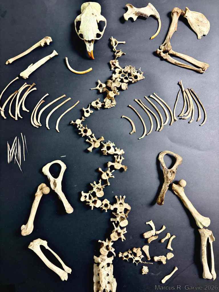
While searching for mammal skulls in the cedar forests of Preservation Park, a large urban park in Guelph, I was fortunate to find an almost complete porcupine skeleton at the base of a tree. The disintegrating quills scattered around the bones made the animal’s identity immediately clear.

The North American porcupine (Erethizon dorsatum) is a large, tree-climbing rodent native to Canada, the United States, and northern Mexico. In this post, I use the references listed above to identify some of the more readily recognizable bones. I didn’t try to identify every bone I found. Here is a picture of my entire collection of bones:
Bones I identified:

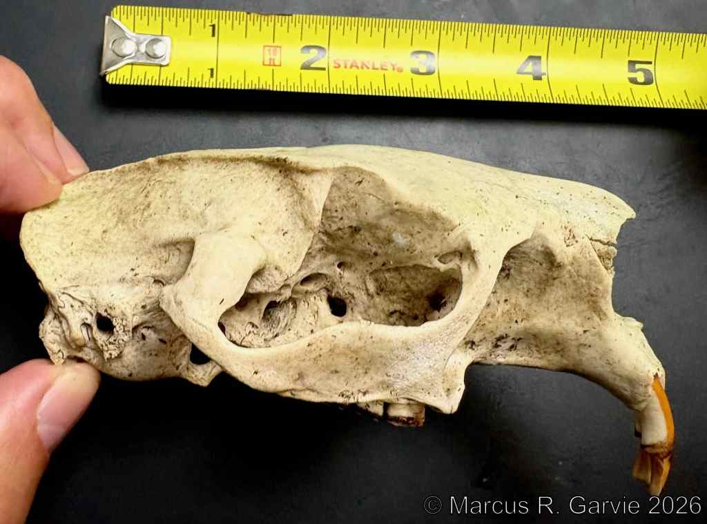
Wow! Look at those temporal ridges.

The teeth keep growing and can be pulled out. They are longer than they look.


The cheek teeth are very distinctive, so it’s worth a closer look. Porcupine molars and premolars are lophodont: their chewing surfaces have enamel ridges and folds that help grind vegetation. Wear creates enclosed enamel loops, which can give the teeth a basin-like appearance.
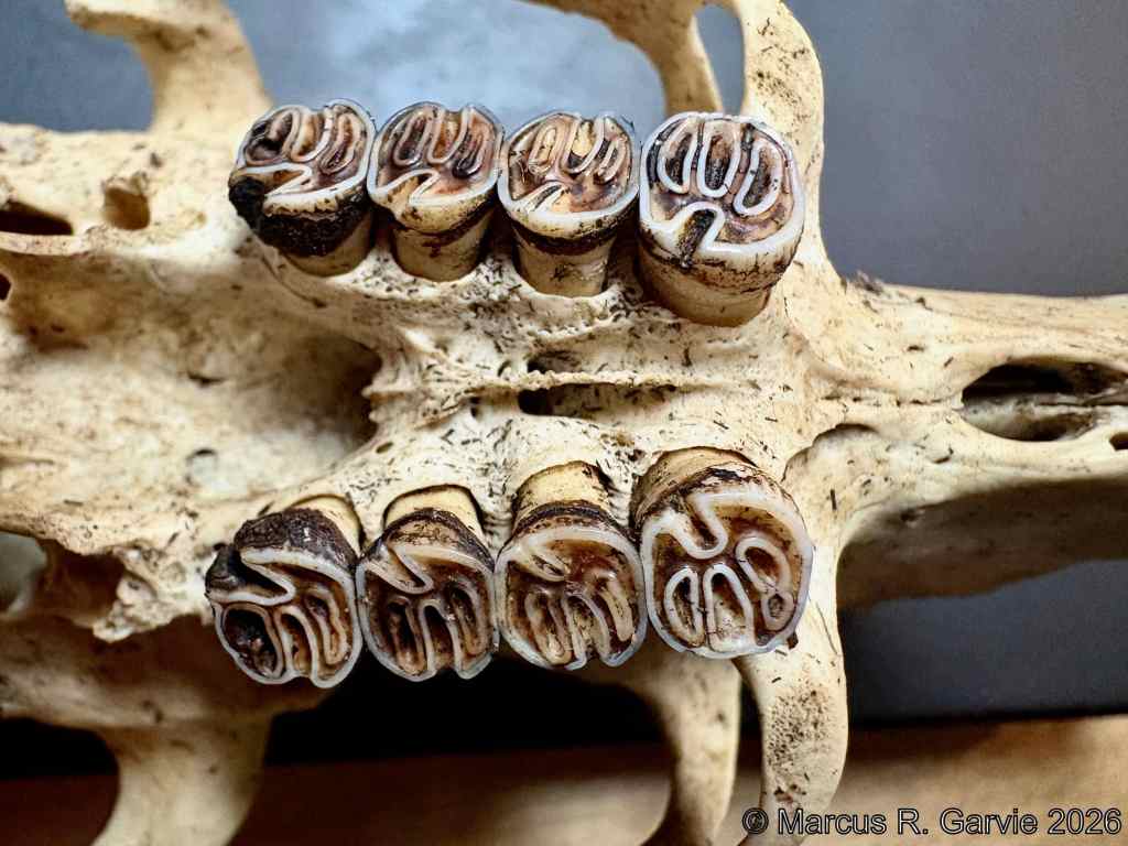
The adult North American porcupine’s dental formula is:
I 1/1, C 0/0, P 1/1, M 3/3 × 2 = 20 teeth
(see [4]). The letters identify the tooth types:
- I = incisors
- C = canines
- P = premolars
- M = molars
Each pair of numbers gives the number of teeth on one side of the mouth, with the upper jaw first and the lower jaw second. Thus, P 1/1 means one upper premolar and one lower premolar on each side. Multiply the combined counts by two to include both sides, giving 20 teeth: four incisors, four premolars, and twelve molars.
I also found one of the mandibles:


I think we can see where this mandible was attached to the left mandible.
And now, some of the main bones.

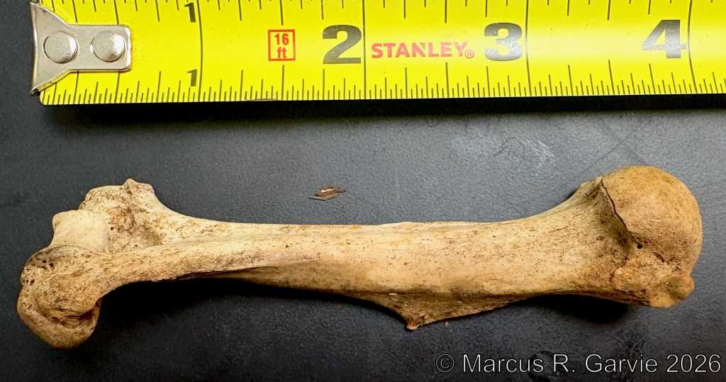

The ulna has a distinctive “U”- shaped indentation.
There are many different-shaped vertebrae, and I did not attempt to distinguish between them.


According to [1], there are 14 ribs on each side of the animal.

It is easy to see which bone connects to the half-pelvis:

Next, we have the tibia (thicker) partially connected to the fibula (thinner):

Finally, we have the Sacrum, some fused vertebrae at the base of the spinal column:

Final Thoughts:
Using the excellent reference [1], I could have identified some of the smaller bones (e.g., I think I see the Patella). But I have run out of time. I used to be daunted by the prospect of learning about the different bones of mammals, but I am beginning to recognize similar features of some of the bones, regardless of the species.

Leave a Reply