Date: Saturday April 25, 2026
Place: Near the town of Mono along the Bruce Trail, close to Mono Cliffs Provincial Park. See the map below:
Context:
This was the first day of the first weekend of the 2026/27 Tracking Apprenticeship Program with Earth Tracks (see https://www.earthtracks.ca/). Most of our observations were signs rather than tracks, and there were so many that I am only discussing those that were new to me, or those for which I want to explore what we found in a little more depth.
General References:
– [1] ‘Mammal Tracks & Sign: A Guide to North American Species’, Mark Elbroch, Stackpole Books (2nd Ed.), 2019
– [2] ‘Tracks & Sign of Insects and Other Invertebrates’, Charley Eiseman and Noah Charney, Stackpole Books (1st Ed.), 2010
Chronological account of some signs:
The first observation is of a mantis ootheca. An ootheca is an egg case. For mantises, it is the foamy mass that the female produces around her eggs. The foam hardens into a protective case, usually attached to a twig, stem, grass blade, fence, or other surface. The eggs overwinter inside it, and the young mantises hatch out later. Looking in [2] the photo below looks most like a European Mantis (Mantis religiosa), an introduced species:
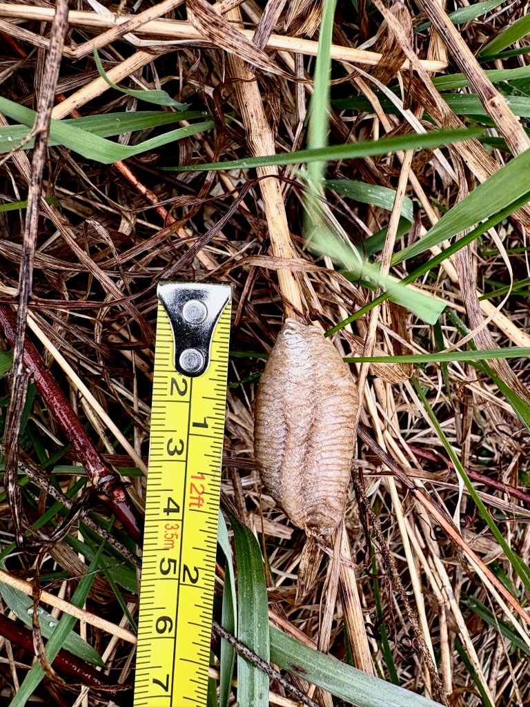
The Chinese mantid (Tenodera sinensis) is also apparently common in southern Ontario, but has a different appearance; see page 15 of [2].
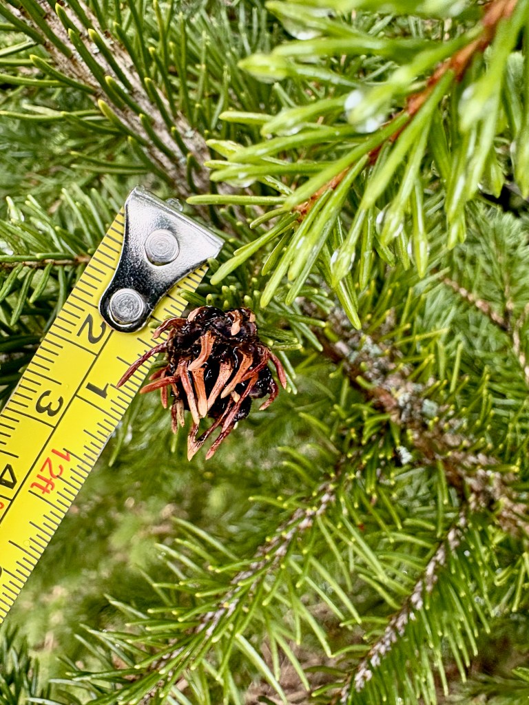
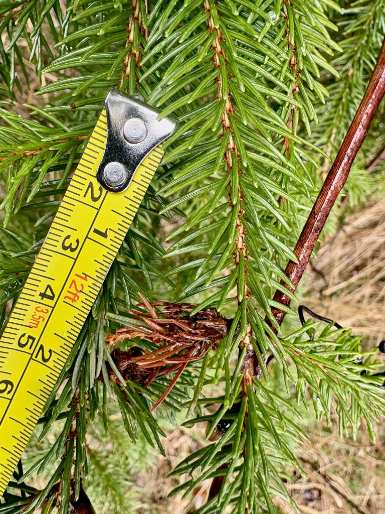
Near where we found the ootheca we also found a gall on a spruce tree. This is the Pineapple-gall Adelgid (Adelges abietis). The adelgids are a group of small, sap-sucking insects related to aphids. Adelgids feed on conifers, and Adelges abietis causes the distinctive pineapple-shaped galls on spruce. Apparently there are several different species of adelgids that produce galls on spruces (see pages 403 – 404 of [2]).
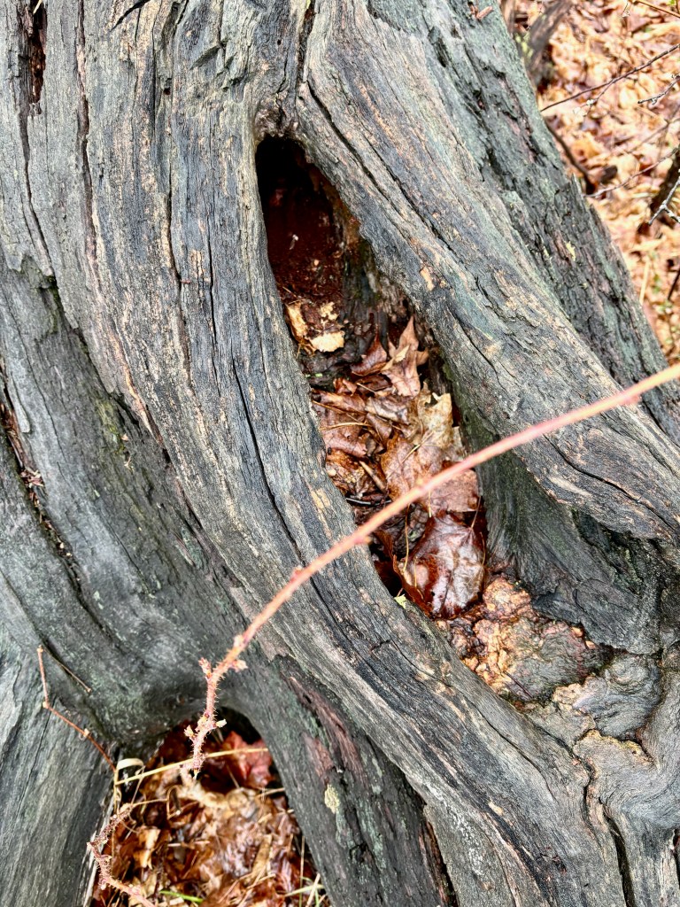
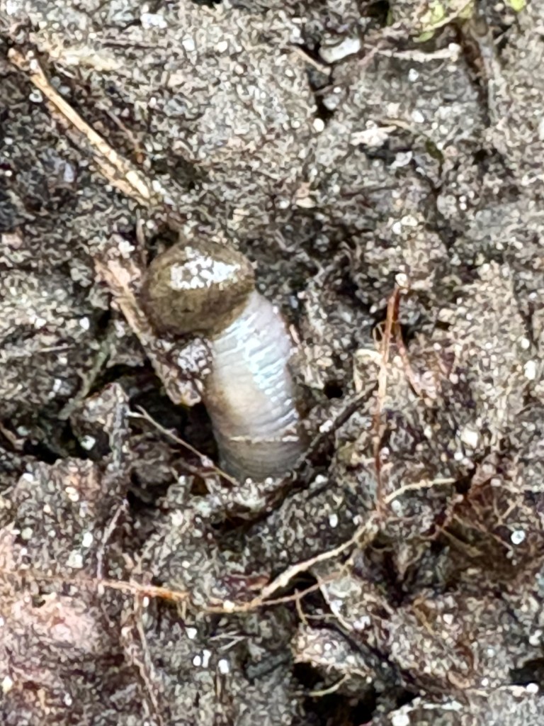
Along the same path, Alexis investigated frass coming out of a hole in a rotting tree. He pointed out some scat that looked fatter than the mouse scat I had seen in my basement. I recognized it as vole scat. The scat was approximately 5 mm long (about 3/16 of an inch), which is in the range for meadow vole scat (see page 143 of [1]).
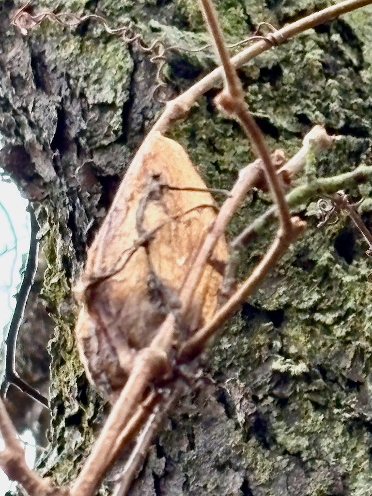
My next observation was of a silk moth cocoon. It was attached quite high on a climbing vine, possibly grape or Virginia creeper, so I was not able to measure it. However, I estimate that it was about 2 – 3 inches long. A silk moth cocoon is a protective, fibrous silk case spun by a silk moth caterpillar around itself, where it pupates and usually overwinters before emerging as an adult moth in spring or early summer. Three common silk moth cocoons are described in
To me the cocoon looks most like the cocoon of a Promethea Moth (Callosamia promethea), but maybe it was too high off the ground?
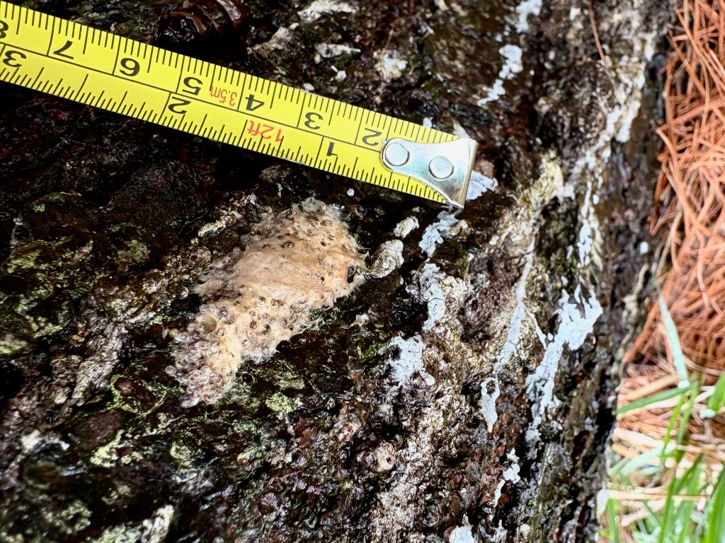
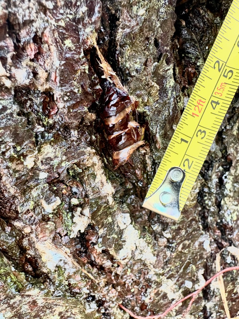
I found both the pupa and egg mass of the Spongy Moth (Lymantria dispar dispar). The pupa is a dark, hardened, segmented case attached to a sheltered surface, representing the resting stage between the caterpillar and the adult moth. Nearby was an egg mass: a tan, fuzzy, oval patch made from hairs from the female moth’s abdomen, which protect the eggs through the winter. These two signs (see pages 22 and 98 of [2]) show different stages in the life cycle of this introduced moth species, whose caterpillars feed on the leaves of many deciduous trees.
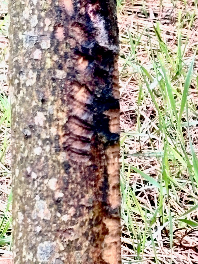
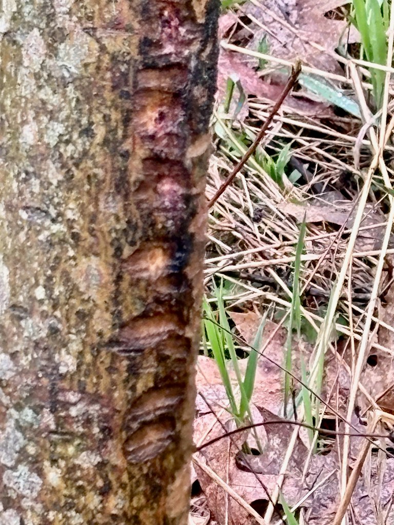
Next, Alexis showed us cambium feeding by White-tailed Deer on the bark of Alternate-leaved Dogwood (Cornus alternifolia). This seemed somewhat unusual, because the deer cambium-feeding sign I have seen before tends to appear as vertical scrapes made by the lower incisors; see page 276 of [2].
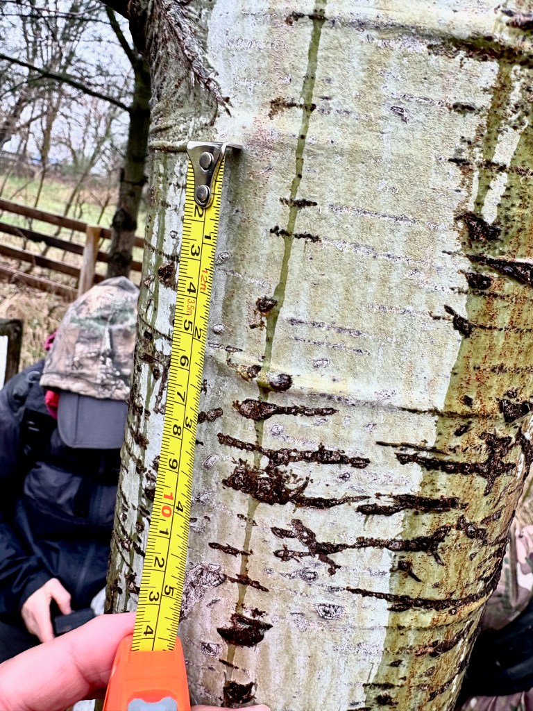
Soon after, we came to a grove of Aspen trees where claw marks from an animal were visible, presumably from spring feeding on the nutritious shoots and early foliage. Alexis suggested that these were made by Raccoons climbing the trees, but I wondered why they could not have been made by Porcupine or Black Bear. I tried searching for the expected distance between claw marks for these two species, but did not find any clear references. A picture of porcupine claw marks on an Aspen tree is shown on page 265 of [1], although the image is not especially clear. Perhaps someone can help me here? I estimate the distance between the claw marks to be about 3/4 inch.
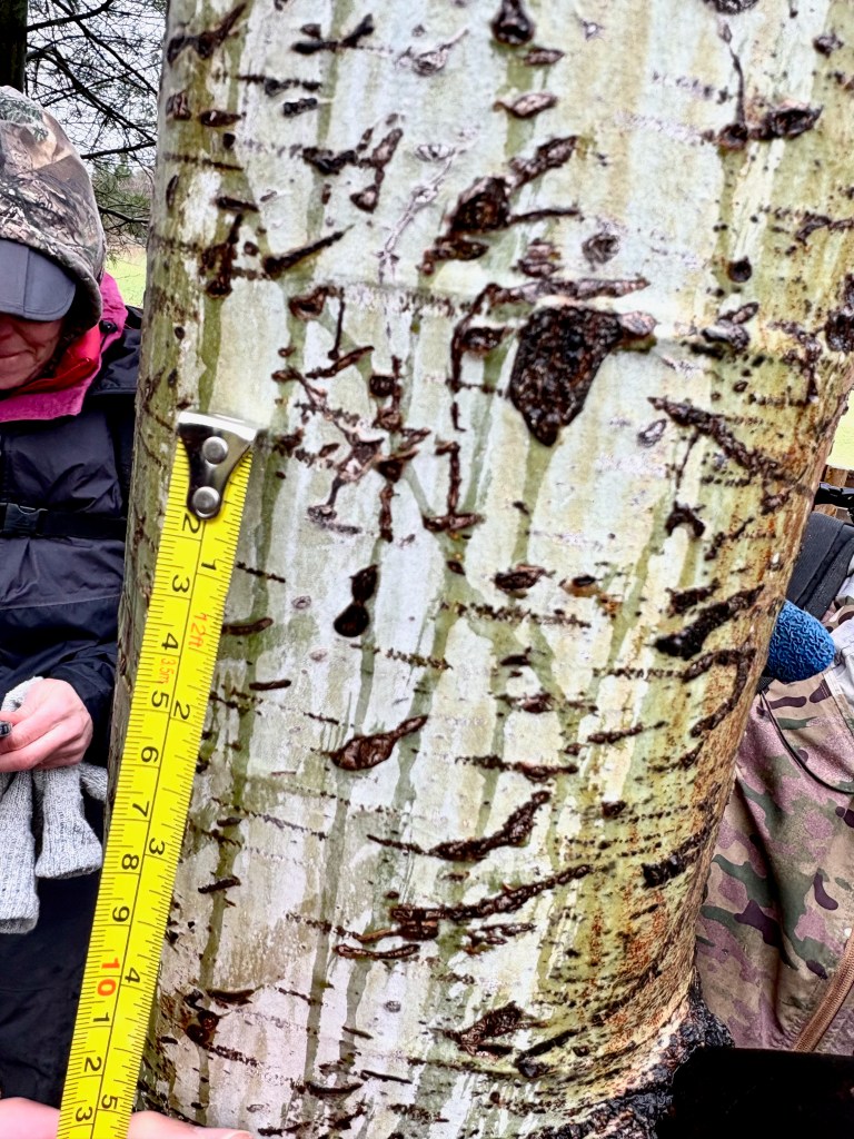
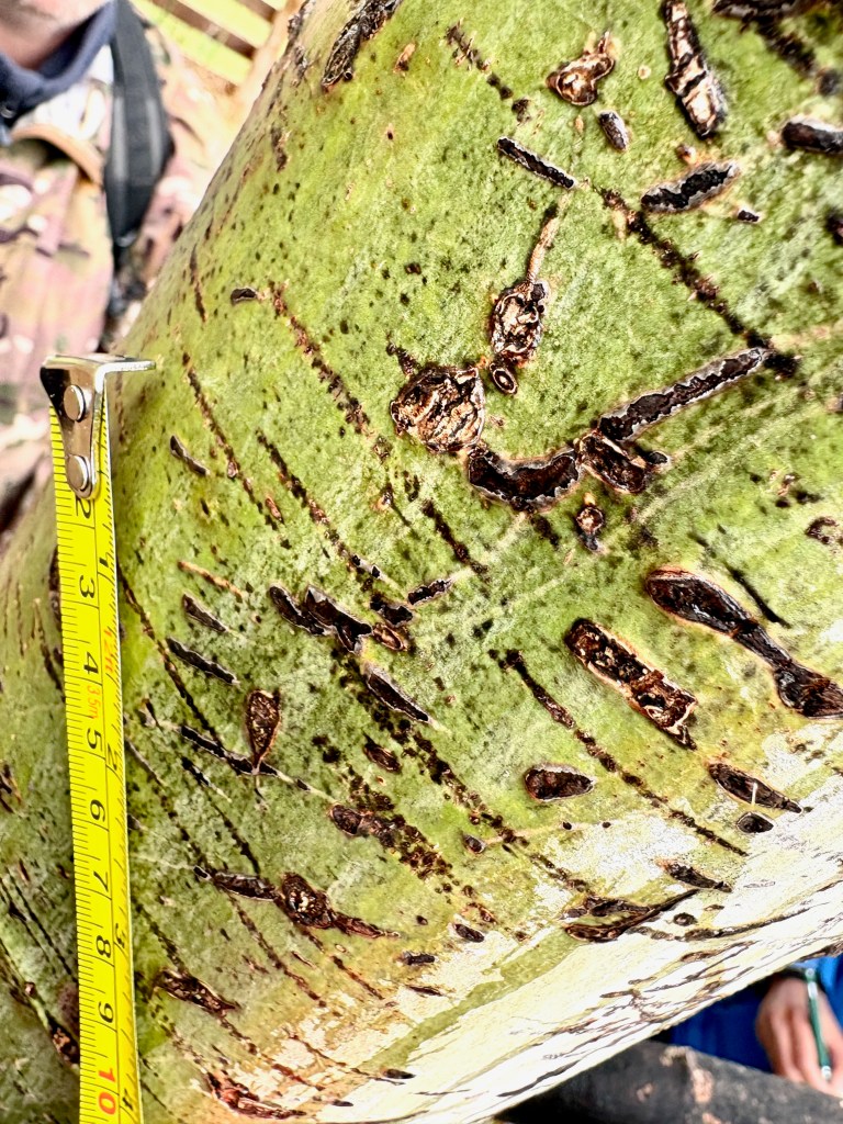
I realized that I can get a rough idea of the spacing between the claws of different animals by looking at the life-size track prints in [1]. This yielded the following rough estimates:
- Porcupine: roughly 1/4 – 1/2 inch
- Raccoon: about 1/2 – 1 inch
- Black Bear: 3/4 – 2 inches
I think raccoon fits best.
In a nearby meadow we also found what appeared to be a mole mound in an area where the soil looked fairly well-drained. That makes Hairy-tailed Mole (Parascalops breweri) a good candidate, since this species occurs in southern Ontario and is associated with mixed, deciduous, and evergreen forests, old fields, roadsides, and other places with lighter, well-drained soils. Star-nosed Mole (Condylura cristata) is also possible in the broader region, but it is more strongly associated with wet meadows, marshes, stream edges, and other poorly drained habitats. Since this mound was not in an obviously wet setting, Hairy-tailed Mole seems like the better fit, although the identification is tentative without seeing the animal or more diagnostic sign.
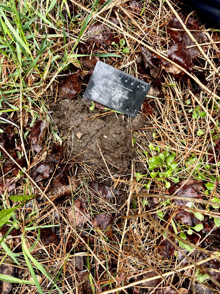
Apparently Hairy-tailed Moles also leave latrines (see page 144 of [1]) so that would be something to look for in the future. There is a good account of this species at https://animaldiversity.org/accounts/Parascalops_breweri/.
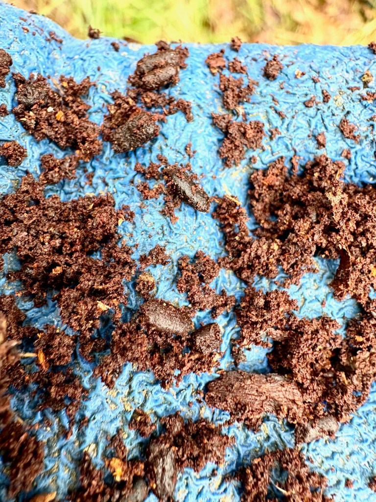
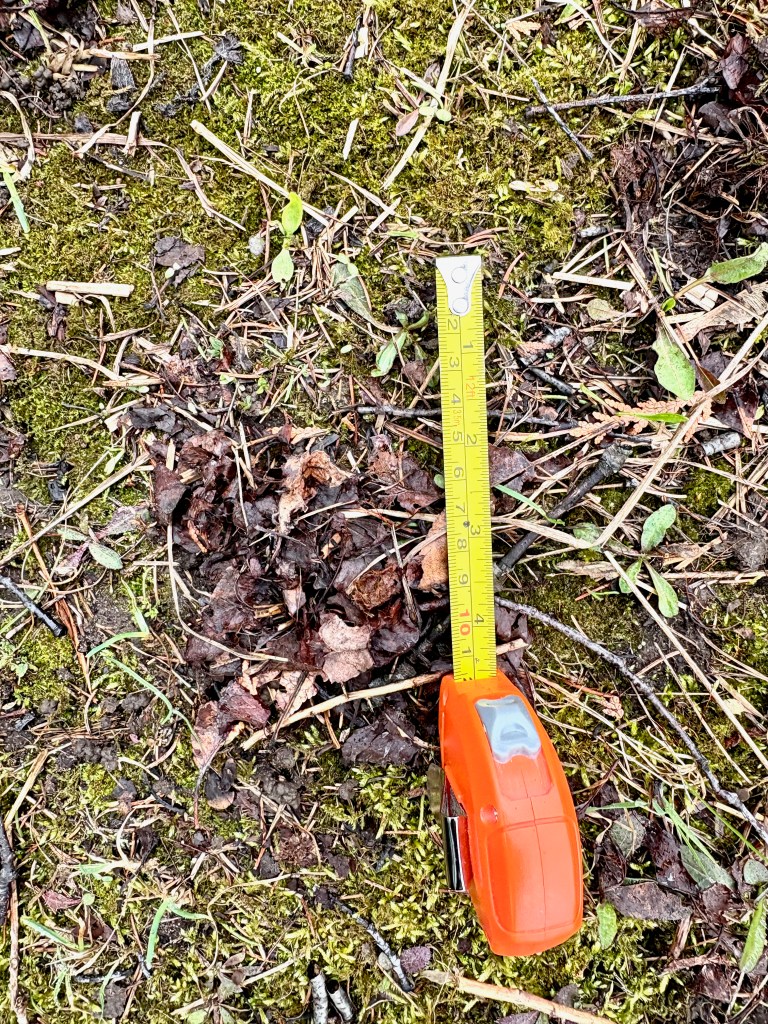
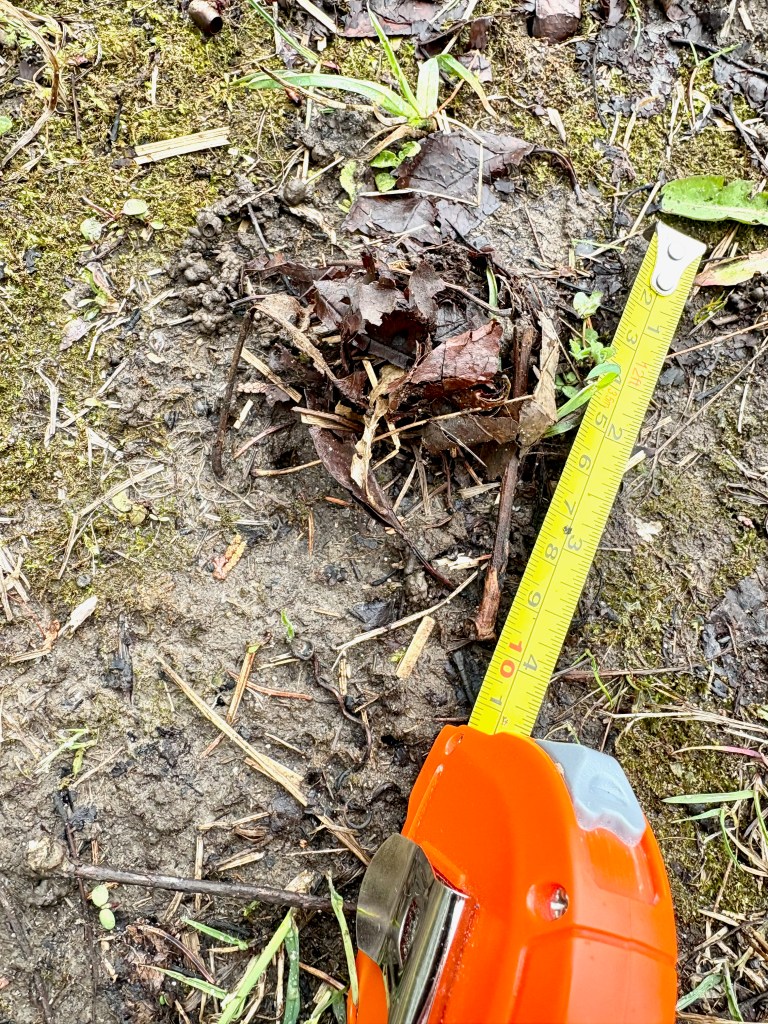
Alexis pointed out small clumped piles of leaves surrounded by bare soil. These appear to be earthworm middens, likely made by common nightcrawlers (Lumbricus terrestris), which live in deep burrows but feed on surface litter. The worms pull leaves and other plant material toward the burrow entrance, where it can be eaten, used to line the burrow, or help protect and regulate conditions inside (see pages 466 – 467 of [2]).
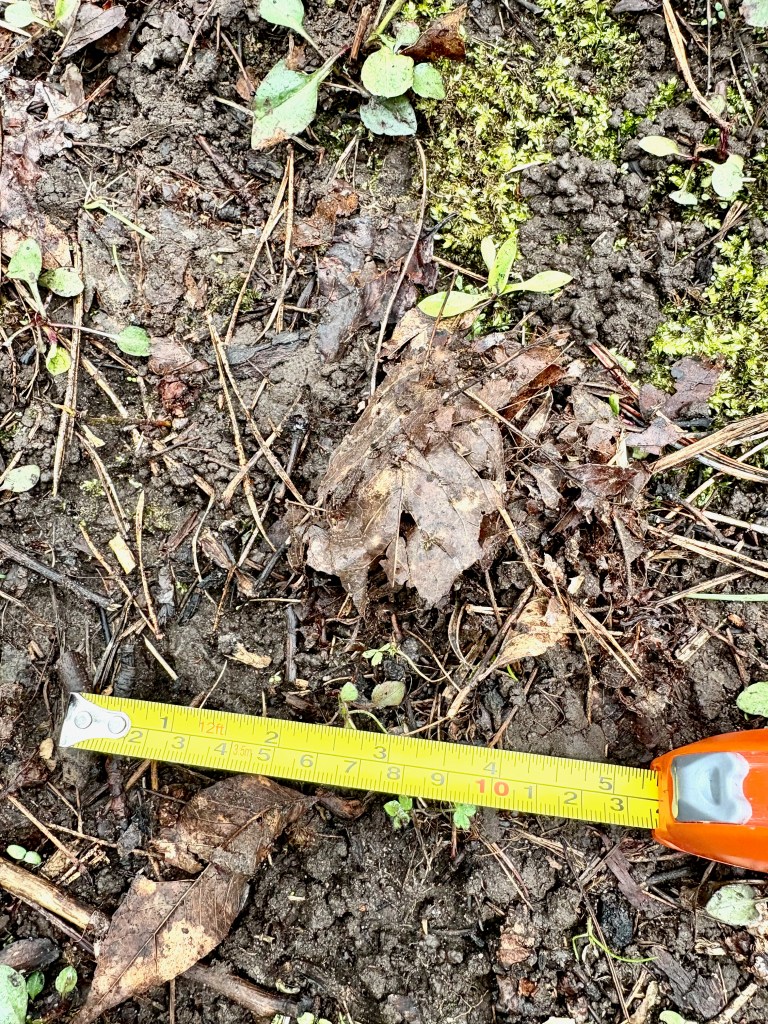
In southern Ontario and much of the Great Lakes region, many earthworms are non-native species introduced from Europe. Although earthworms can be useful in gardens and farms, in forests they can have harmful effects by rapidly breaking down and mixing leaf litter into the soil. This can leave the forest floor with only a thin layer of fresh leaves instead of a rich, layered blanket of decomposing litter. Many native forest plants and small animals depend on that litter and duff layer, so heavy earthworm activity can lead to a sparser, more disturbed understory, sometimes favouring only a few tolerant plants, such as certain ferns, sedges, or introduced Eurasian species (see page 444 of [2]).
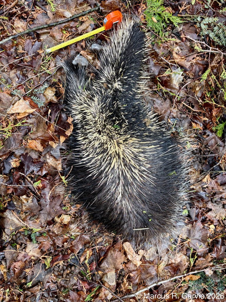
Later, when we came to the top of a ridge in the forest, we found a dead porcupine in very good condition. We wondered whether it might have died from a fall. North American Porcupines are strong climbers, but they are known to fall from trees occasionally, and studies of porcupine skeletons have found that roughly 30% of the animals examined had healed fractures, suggesting that falls are not unusual and that many porcupines survive them. In this case, the idea seemed plausible because a little farther up the hill we found a small collection of quills on the ground, which may have marked the spot where it fell. Of course, without a closer examination there is no way to know for certain, but a fall seems like one possible explanation. Here are some references that support what I said.
- Roze, U., Locke, D. C., & Vatakis, N. (1990). Antibiotic properties of porcupine quills. Journal of Chemical Ecology, 16, 725–734. See https://pubmed.ncbi.nlm.nih.gov/24263588/
- Alaska Department of Fish and Game. “North American Porcupine (Erethizon dorsatum) Species Profile.” See https://www.adfg.alaska.gov/index.cfm?adfg=northamericanporcupine.main
This dead porcupine provided an excellent opportunity to study its front and hind feet and relate them to the typical tracks they leave. The following track prints are from [1]:
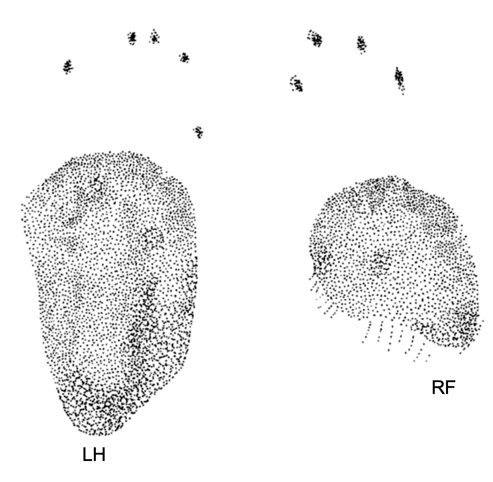
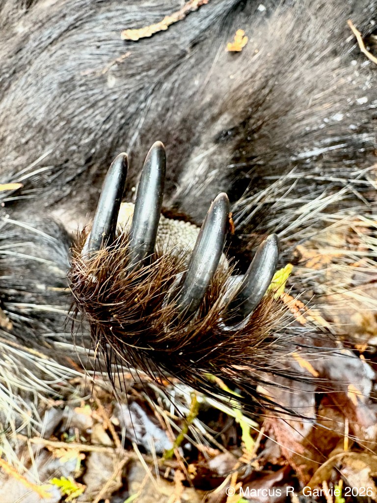
Let’s look at the hind feet first:
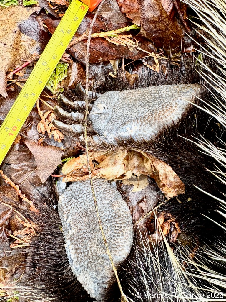
Notice the pebbly texture of the feet, five toes on each foot, the dropped toe on the inside, and the long toes that are typically not seen in the track because the claws raise them above the ground. The metatarsal (palm) and heel pads are fused into a single oval pad. It also looks like the pad is longer on the outside of each foot.
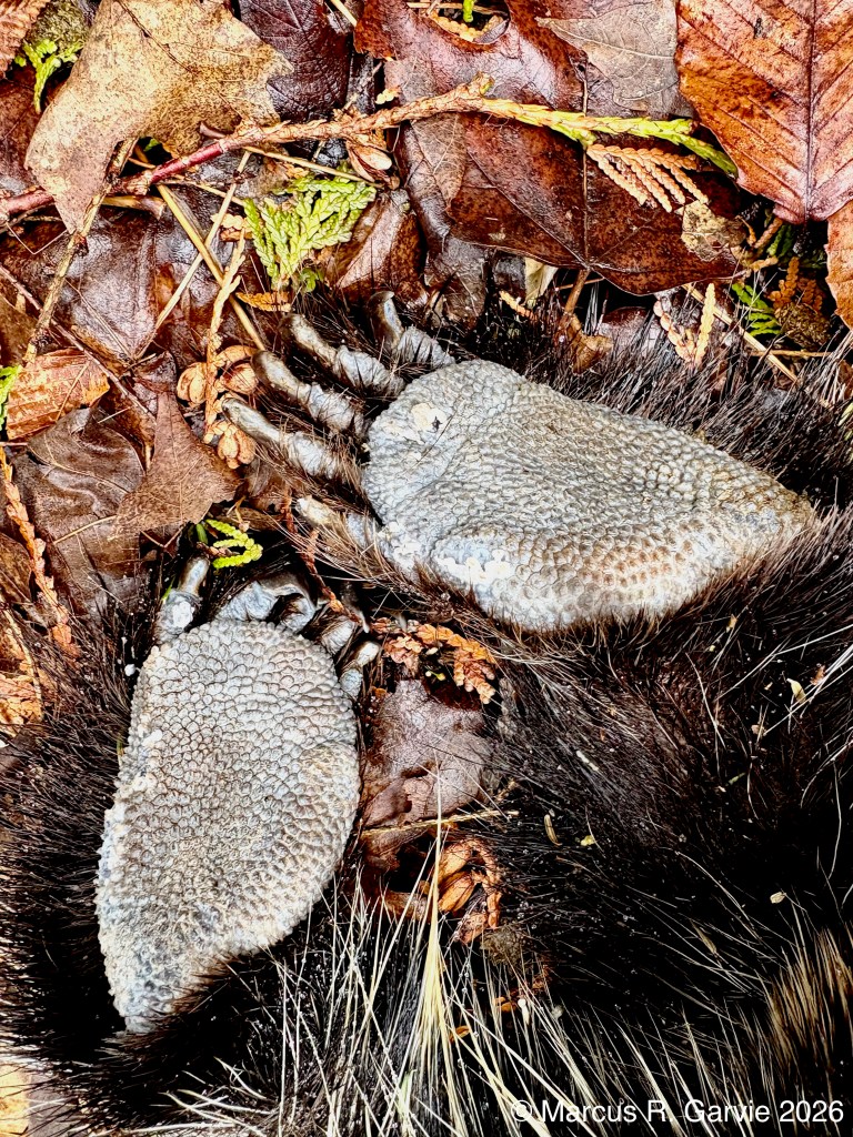
Here are photos of the front feet:
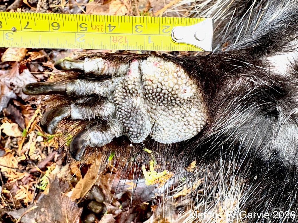
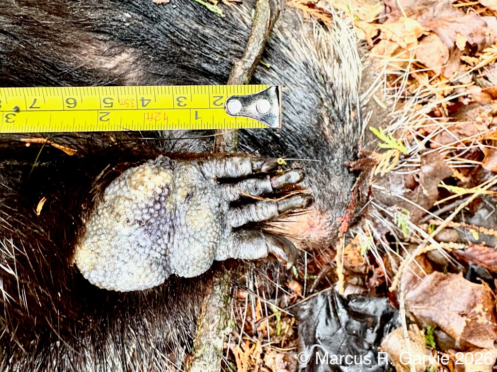
There are two fused metacarpal (palm) pads. Am I seeing a ‘crease’ in the foot separating the palm pad from the heel pad? This isn’t mentioned in the description given by [1] (see page 578). Look at those claws!
I also took a closer look at the quills and hair of the porcupine:
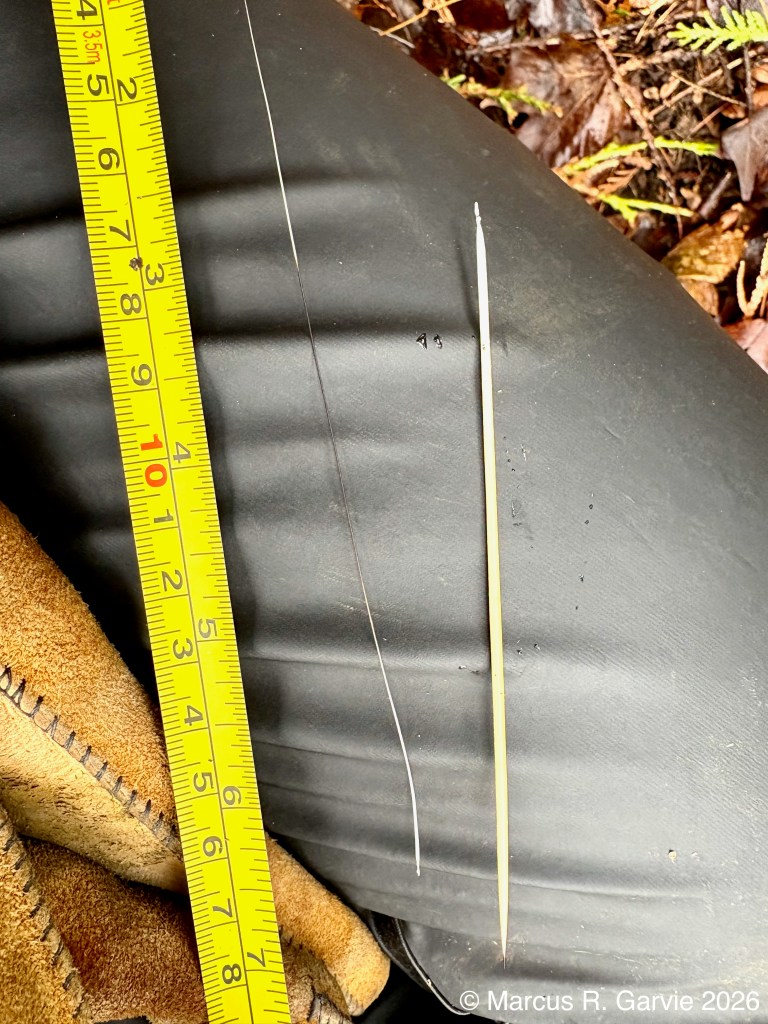
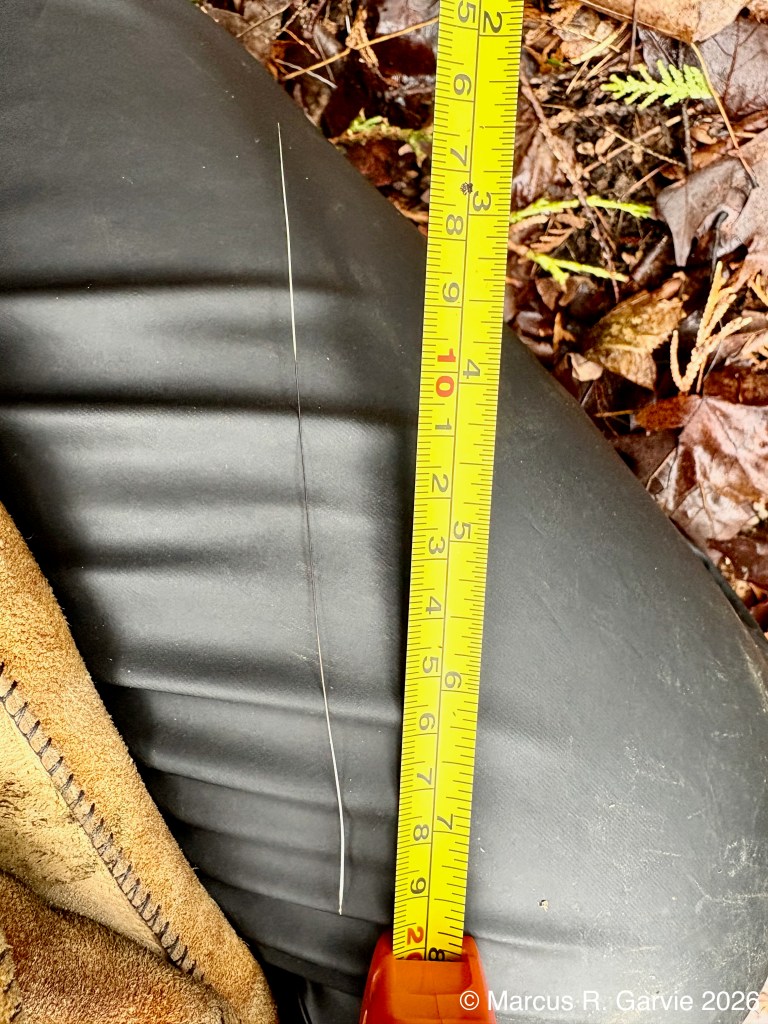
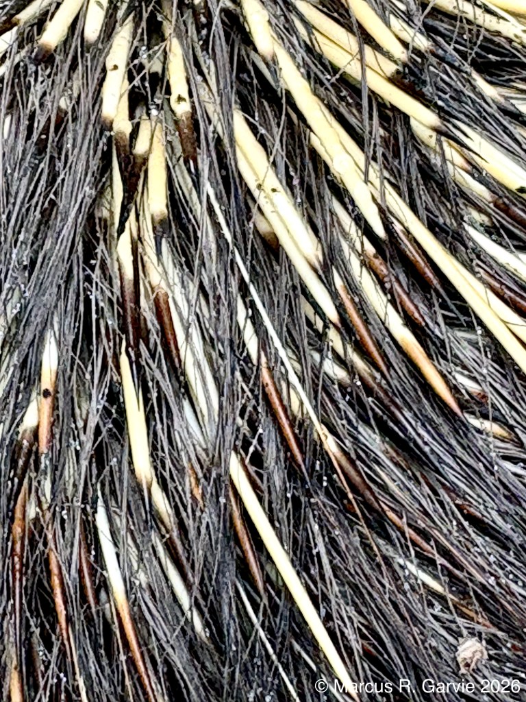
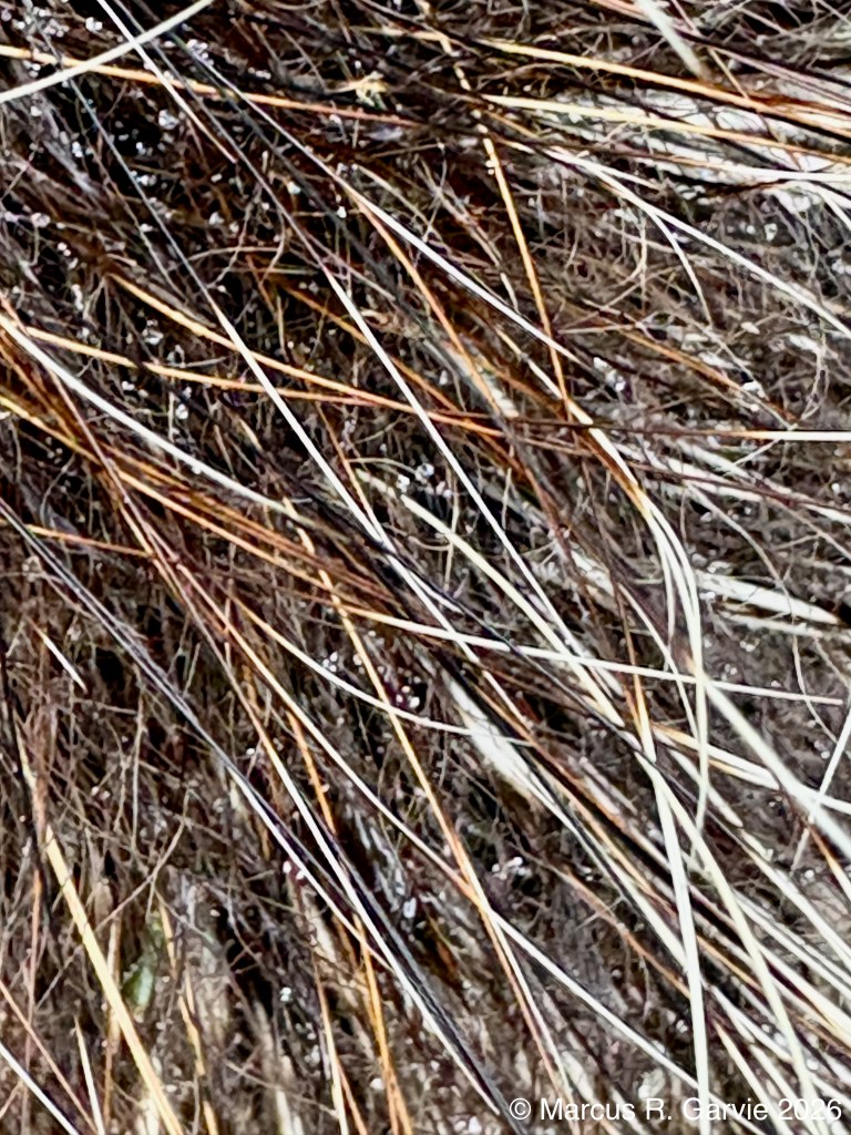
I noticed that the hair is multi-coloured, with some strands almost 5 inches long. The quill I pulled out was 4 1/2 inches long. The quills are interspersed with the fur, but some areas of the body, such as the back and tail, have a much greater density of quills, presumably because these are the areas where the animal needs the most protection.
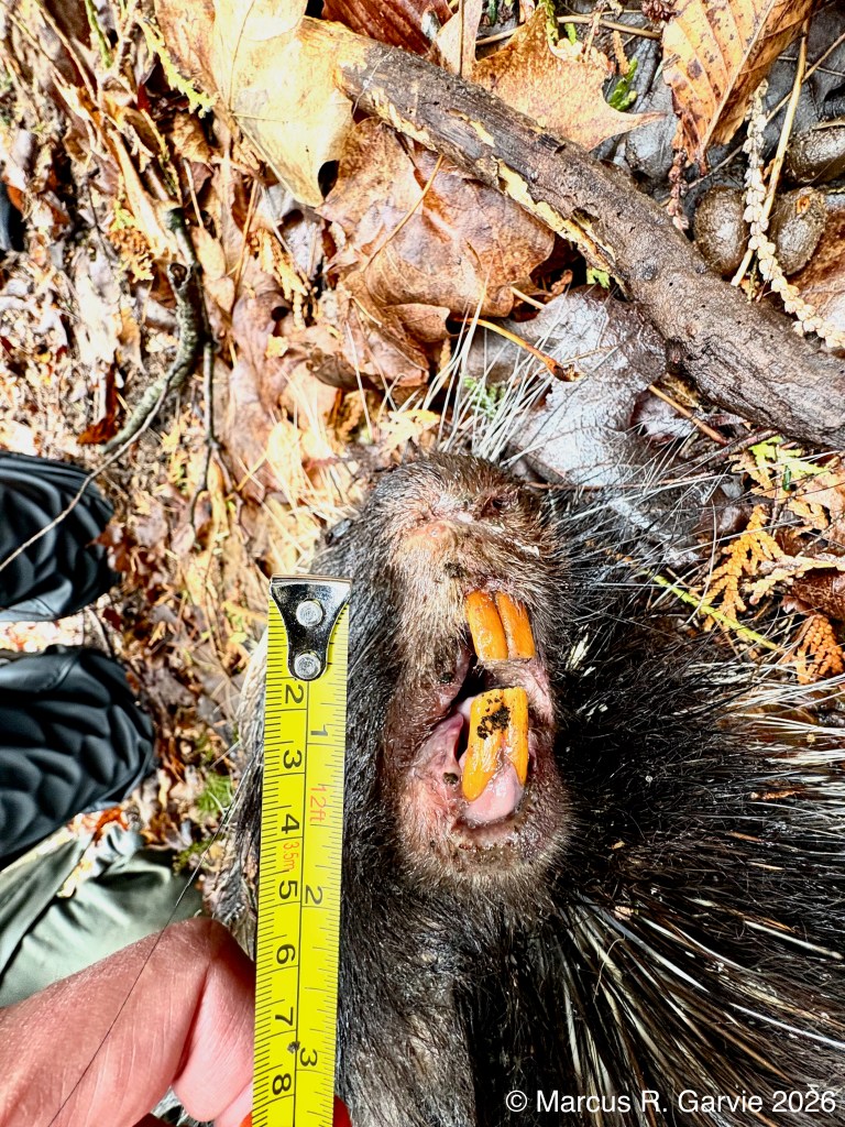
At another location we also found the skull and some bones of a porcupine:
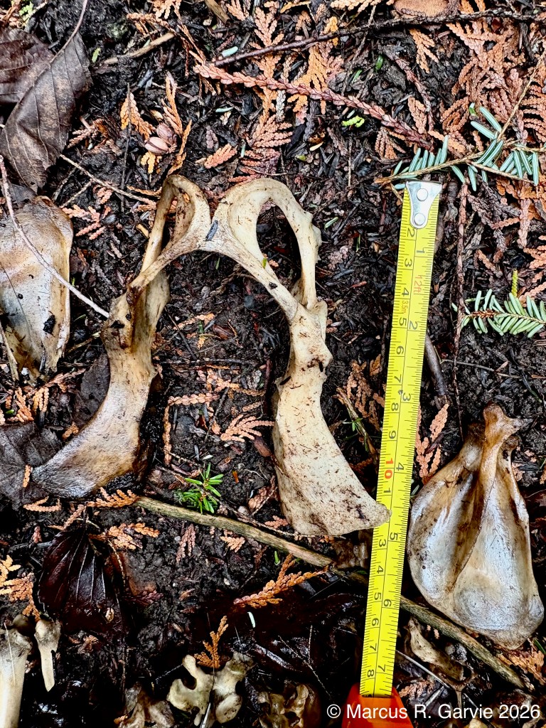
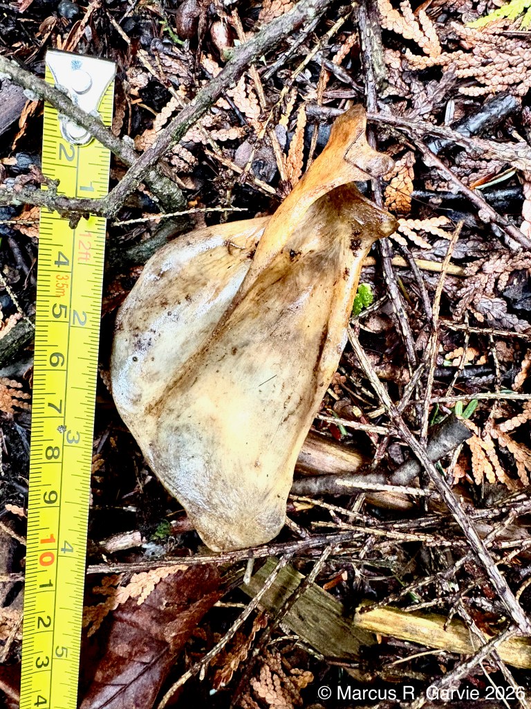
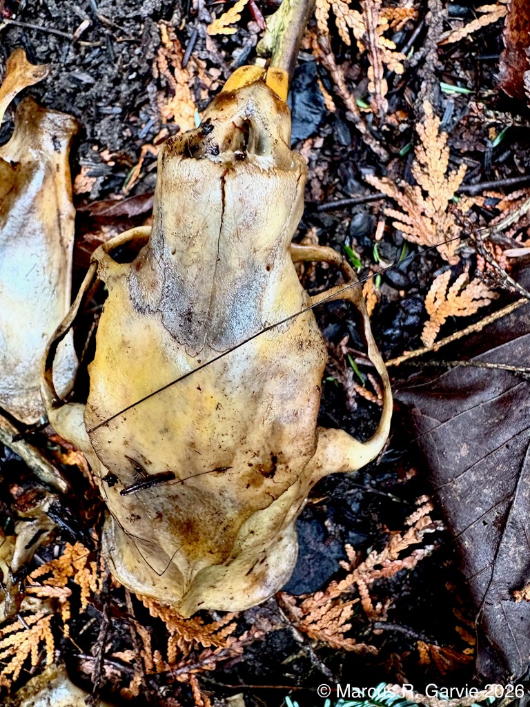
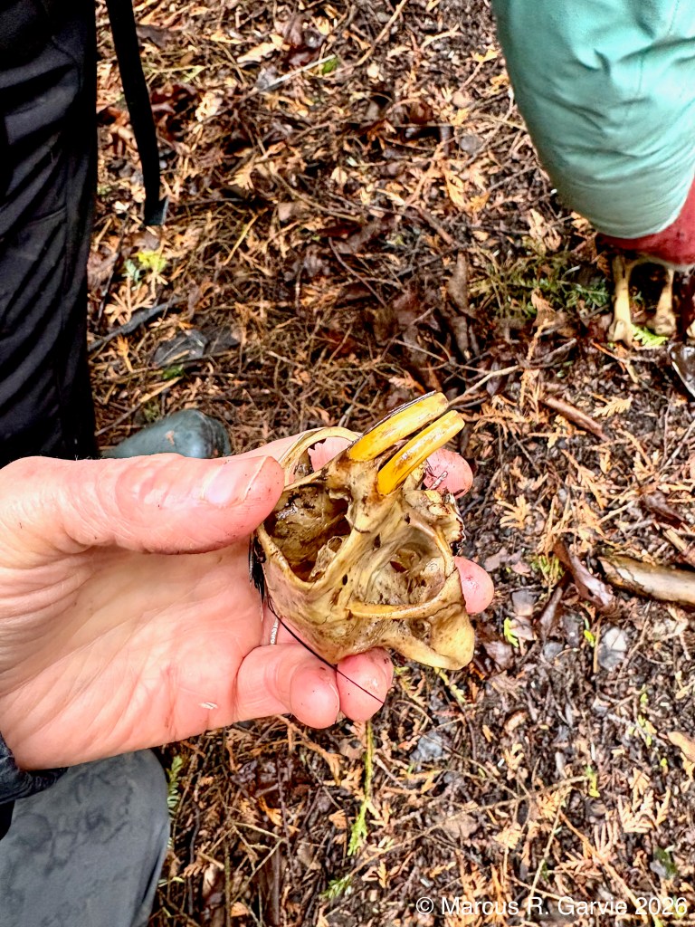
These photos show, respectively, the pelvis, the scapula, and two views of the skull of a porcupine. Like all rodents, porcupines have continuously growing incisors that are kept worn down by gnawing. In the skull, the incisors are surprisingly long and curve far back into deep sockets in the jaws. In this specimen, I was able to slide the incisors partly out, making them appear longer; this is not true “retraction,” but rather reflects how much of the tooth is normally hidden inside the jaw.
A fellow tracker pointed out that the porcupine scapula has a large, fin-like ridge that would have served as an attachment point for strong shoulder and upper-back muscles. In other words, the ridge is part of the framework those muscles pulled against when the animal moved its front legs and braced its body. That made sense to me, since porcupines are such capable climbers.
At the end of our tracking day we also found a raccoon skull:
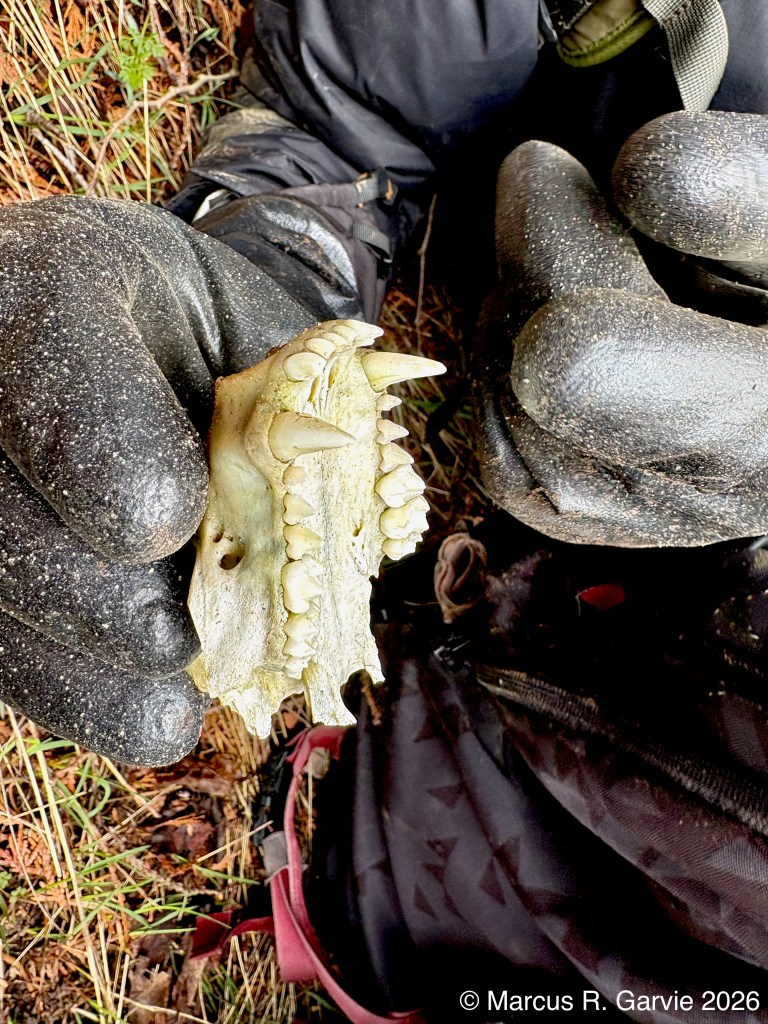
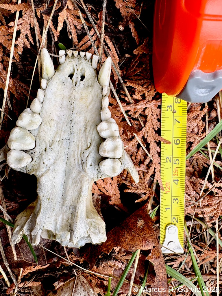
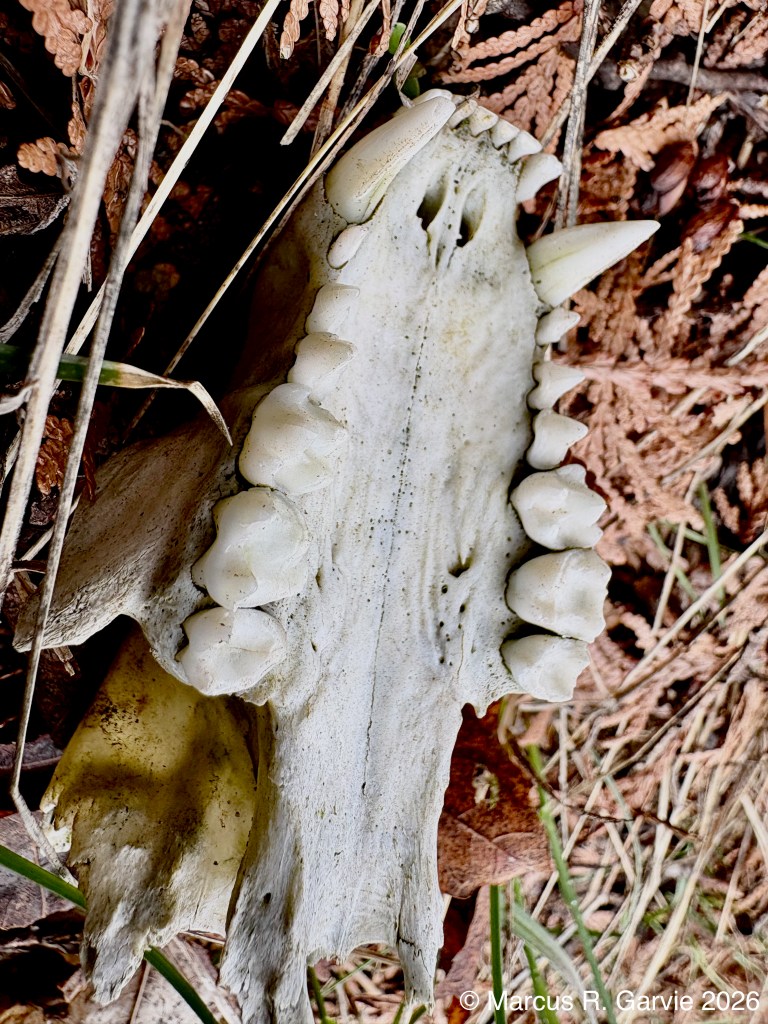
These photos show the underside of the upper part of a raccoon skull, with the broad hard palate running down the middle and the upper tooth rows on either side. At the front are the two large upper canines, and between them are the six small front incisors: three on the left and three on the right. Behind each canine are the cheek teeth, arranged as 4 premolars + 2 molars = 6 cheek teeth on each side. The premolars are the teeth just behind the canine; they are generally smaller and more pointed. The molars sit farther back and are broader and flatter for grinding. A useful basic reference for these skull features is the Maryland DNR’s simplified furbearer skull key, though I am still at the very start of my journey in learning how to understand animal bones and skulls, see https://dnr.maryland.gov/wildlife/documents/skullkey.pdf .

Leave a comment